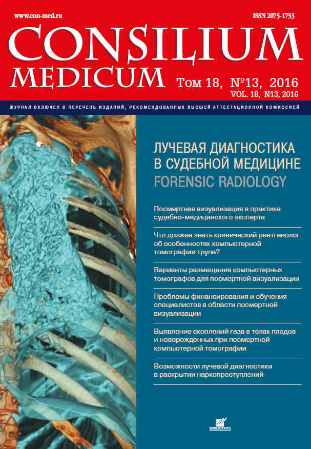
Том 18, № 13 (2016)
- Год: 2016
- Выпуск опубликован: 20.12.2016
- Статей: 6
- URL: https://consilium.orscience.ru/2075-1753/issue/view/4820
Весь выпуск
Статьи
Реальные возможности посмертной лучевой диагностики в практике судебно-медицинского эксперта
Аннотация
Цель исследования. В обзоре проанализированы результаты исследований по проблеме использования компьютерной томографии (КТ) и магнитно-резонансной томографии (МРТ) в судебно-медицинской экспертизе (СМЭ) взрослых лиц. Целью настоящего обзора является ознакомление судебно-медицинских экспертов и рентгенологов с ситуациями, наиболее распространенными в посмертной визуализации, а также с ее сильными и слабыми сторонами. Материал и методы. Использованы основные интернет-ресурсы: Российская научная электронная библиотека (elibrary.ru), Embase, Medline, Web of Science, Cochrane database. Результаты и выводы. Методы посмертной визуализации в СМЭ находятся на стадии активного изучения и формирования доказательной базы. «Золотым стандартом» посмертной диагностики остается традиционная аутопсия. Но роль томографических методов исследования постоянно растет. Для практических целей СМЭ трупов взрослых в большей степени подходит КТ. В ряде случаев следует комбинировать с аутопсией разные лучевые методы диагностики - КТ, КТ-ангиографию и МРТ. Посмертная лучевая диагностика может оказать большую помощь в визуализации механических повреждений, а также в установлении причины в ряде случаев скоропостижной смерти. Посмертная визуализация может быть востребована и в некоторых других распространенных в практике СМЭ ситуациях: механической асфиксии, утоплении, действии высокой и низкой температуры, исследовании гнилостно измененных и неопознанных трупов, выявлении инородных тел.
Consilium Medicum. 2016;18(13):9-25
 9-25
9-25


Выявление скоплений газа в телах плодов, мертворожденных и умерших новорожденных при посмертном компьютерно-томографическом исследовании
Аннотация
Посмертная компьютерная томография (КТ) требует проведения дифференциальной диагностики посмертных и прижизненных патологических процессов. Наиболее актуально данный вопрос стоит в отношении выявления скоплений газа, поскольку традиционное патологоанатомическое исследование не позволяет в полной мере выявить наличие внутриорганного или внутрисосудистого газа за исключением выраженной воздушной эмболии. Цель исследования. При помощи посмертной КТ изучить особенности локализации скоплений газа в органах и тканях погибших плодов, мертворожденных и умерших новорожденных. Материалы и методы. Проведана посмертная КТ 110 плодов, мертворожденных и умерших новорожденных. Все наблюдения были разделены на пять групп. Две группы составили плоды после самопроизвольного (1-я группа, n=9) или индуцированного аборта (2-я группа, n=41) на 14-21-й неделе гестации. Две группы составили тела мертворожденных, погибших на гестационном сроке 25-39 нед антенатально (3-я группа, n=15) с давностью внутриутробной гибели от 14 ч до 2 нед или интранатально (4-я группа, n=3). В 5-ю группу вошли тела 42 новорожденных, рожденных на сроках 24-40 нед и умерших в возрасте от 6 ч до 166 дней. После КТ-исследования проводили патологоанатомическое вскрытие с последующим анализом гистологических препаратов тканей и органов. Результаты. Реже всего скопления воздуха наблюдались при посмертной КТ тел плодов после индуцированных (14,6%) и самопроизвольных (11,1%) абортов. Чаще всего газ визуализировался в случаях интранатальной смерти и у умерших новорожденных. На томограммах тел новорожденных, погибших интранатально, газ визуализировался во всех 3 наблюдениях. Только у 5 (11,9%) умерших новорожденных скопления газа не определялись. Выводы. Посмертная КТ является более эффективным методом выявления скоплений газа по сравнению с аутопсией, однако не может в полной мере являться альтернативой традиционному аутопсийному исследованию, позволяющему проводить комплексное макроскопическое и микроскопическое исследование органов и тканей.
Consilium Medicum. 2016;18(13):26-33
 26-33
26-33


Преимущества и недостатки вариантов размещения компьютерных томографов для посмертной визуализации (опыт специалистов Великобритании)
Аннотация
В статье показаны преимущества и недостатки трех вариантов размещения компьютерных томографов - КТ (непосредственно в морге, на некотором удалении от морга - мобильный КТ-сканер, использование клинических КТ-сканеров) и организации в них посмертной томографической визуализации (PCSI). Подробно по каждому из вариантов рассмотрены проблемные вопросы по доставке трупов, финансированию PCSI-исследований и порядку работы медицинского персонала. На примере функционирования национальной службы Великобритании (NHS UK) показана необходимость разного использования сканирующего оборудования с учетом уникальных особенностей работы каждого морга (отдела патологии госпиталя) страны. Предпочтительной моделью организации рабочего процесса для патологов Великобритании и службы коронеров, а также для родственников умерших является модель размещении сканера непосредственно в морге (вариант 1). Значительные преимущества данного варианта выделяют его среди остальных за счет максимальной эффективности и высокого качества проводимых исследований, а также возможности широкого обучения медицинского персонала (врачи, лаборанты), осуществления эффективного контроля качества клинической диагностики и лечебного процесса, развития новых научных исследований. В силу значительных капиталовложений в масштабах страны целесообразно использование мобильных сканеров (вариант 2) или оптимизации работы клинических сканеров для PCSI-исследований (вариант 3).
Consilium Medicum. 2016;18(13):34-37
 34-37
34-37


Что должен знать клинический рентгенолог об особенностях компьютерной томографии трупа?
Аннотация
В обзоре проанализированы исследования по проблеме посмертной компьютерной томографии (КТ) в судебно-медицинской экспертизе трупа. Основная цель обзора - показать клиническим рентгенологам особенности КТ трупа. При подготовке обзора были использованы основные интернет-ресурсы: научная электронная библиотека (Elibrary), SciVerse (ScienceDirect), Scopus, PubMed и Discover. В обзор включены статьи, в которых обсуждались особенности посмертной КТ, важные для понимания проблемы клиническими рентгенологами, не сталкивающимися в своей повседневной деятельности с посмертной визуализацией. Ранние и поздние трупные изменения, такие как окоченение, аутолиз, гнилостные и другие посмертные процессы, в значительной степени меняют «норму» при КТ-визуализации. Рентгенологи при интерпретации КТ-изображений трупа должны учитывать наиболее часто встречающиеся артефакты, к которым приводят: посмертные свертки крови в полостях сердца и крупных сосудах, аспирация содержимого желудка в воздухоносные пути, эзофаго - и гастромаляция, трупные гипостазы во внутренних органах, нарушение дифференцировки между серым и белым веществом головного мозга, гнилостные газы в сосудах, органах и тканях и многие другие. Посмертная визуализация требует специальной подготовки рентгенологов по судебной медицине и имеет особенности лучевой картины, свойственные только трупу. При отсутствии специальных знаний даже очень опытные и грамотные клинические рентгенологи могут допустить серьезные диагностические ошибки при интерпретации посмертных КТ-изображений.
Consilium Medicum. 2016;18(13):38-47
 38-47
38-47


Проблемы финансирования и обучения специалистов в области посмертной томографической визуализации в Великобритании
Аннотация
В статье описаны возможные варианты выполнения аутопсий в Великобритании с использованием различных дополнительных методов исследования, включая посмертную томографическую визуализацию (postmortem cross-sectional imaging - PCSI). Показано, что официально утвержденной программы подготовки специалистов по PCSI не существует и такая совместная работа ведется в некоторых центрах Великобритании в инициативном порядке. Доказывается необходимость разработки собственных национальных стандартов и учебных программ в рамках каждой профессии с созданием соответствующих систем аудита (проверки, инспекции) и внешнего контроля качества PCSI. Отмечено, что выполнение и хранение томографических данных осуществляется главным образом в учреждениях Национальной службы здравоохранения Великобритании с оплатой работы специалистов по второй категории (аналог выполнения судебно-медицинской экспертизы). Во избежание стагнации и торможения развития данной работы крайне важно, чтобы в основе данного нового научного направления лежала сильная академическая исследовательская стратегия, подкрепленная устойчивым финансированием, осуществляемым на национальном уровне. Показана важность полноценного материального обеспечения PCSI и разработки собственных стандартов, доказывающих возможность использования PCSI в качестве альтернативы для классической аутопсии.
Consilium Medicum. 2016;18(13):48-51
 48-51
48-51


Возможности лучевой диагностики в выявлении наркотических средств, перевозимых контейнерным способом в полостях тела человека (наркокурьера)
Аннотация
В статье обсуждаются возможности лучевых методов исследования - рентгенографии, компьютерной томографии (КТ), магнитно-резонансной томографии (МРТ) и сонографии в поиске и обнаружении наркотических средств (НС), перевозимых контейнерным способом в полостях тела человека (наркокурьера). В обзоре приводятся исследования (по состоянию на сентябрь 2016 г.), показывающие преимущества КТ перед стандартной рентгенографией брюшной полости при поиске контейнеров с НС в желудочно-кишечном тракте. В настоящее время КТ, в том числе низкодозная, может считаться методом выбора для поиска контейнеров с НС в полостях тела человека. КТ следует использовать как для первичной диагностики, так и для повторного исследования после извлечения контейнеров с НС. МРТ и сонография, несмотря на ряд очевидных преимуществ, в том числе отсутствие лучевой нагрузки, имеют ограниченное применение.
Consilium Medicum. 2016;18(13):52-58
 52-58
52-58


